
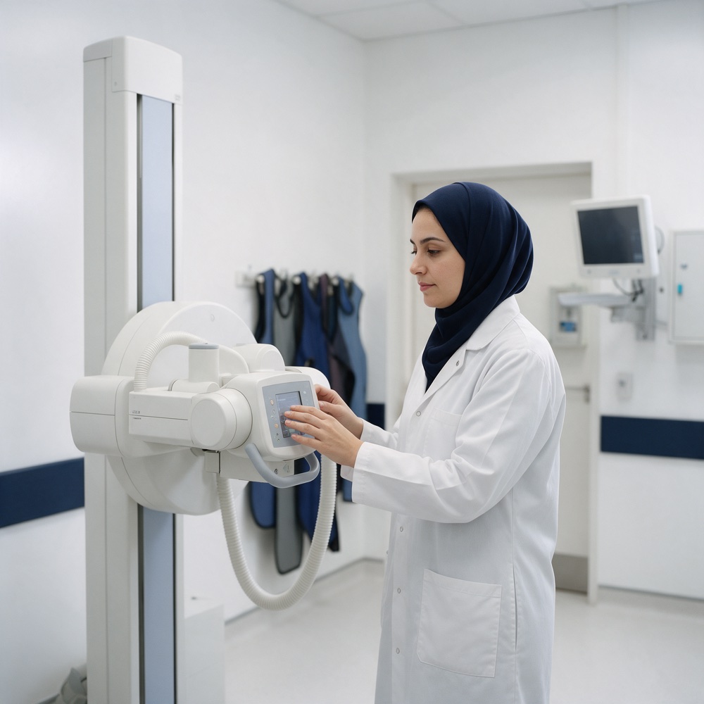
X-ray imaging of the chest, bones and joints — one of the fastest and most requested exams for injuries, chest pain and follow-up after fractures.
A complete panoramic image of the jaws and teeth in a single shot, showing wisdom teeth, roots and the surrounding bone — the first step before many dental procedures.
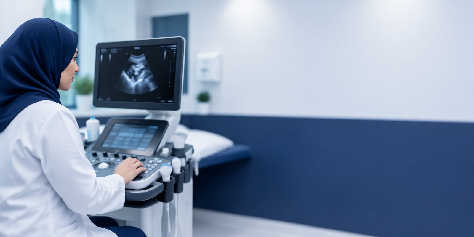
An ultrasound exam that assesses the liver, gallbladder, kidneys, pancreas, spleen and bladder — often requested for recurrent abdominal pain, bloating and changes in lab results.
Monitoring the baby’s growth, heartbeat, position and the amount of fluid around it throughout pregnancy, in coordination with the obstetrician-gynecologist inside the branch.
A more precise scan that reviews the baby’s organs one by one — heart, brain, kidneys, spine and limbs — looking for any congenital abnormality. It is usually done once during pregnancy at a specific time.
A moving three-dimensional image that clearly shows the baby’s features. A scan for reassurance and for seeing the features, with an ideal window when the image is at its clearest.
Radiology and Diagnostic Imaging Specialist — she supervises the department’s exams and the reading of their results at the Al-Taawun Branch, with a female team for women’s and pregnancy exams.
The department is not limited to pregnancy and abdominal scans. These exams are done daily at the Al-Taawun Branch, and you can come directly to the Radiology Department to have them without needing a clinic appointment.
Ultrasonography — Thyroid
Assesses the size of the gland and its nodules — requested for an underactive or overactive thyroid and changes in lab results.
Ultrasonography — Breast
An ultrasound exam of one or both sides — for routine screening or when you feel a lump or pain.
Doppler — Limbs
Examines blood flow in the veins and arteries of one or both limbs — requested for varicose veins, leg swelling and suspected clots.
Doppler U/S Carotid System
Assesses blood flow in the neck arteries that supply the brain — an essential part of following up heart and arterial diseases.
Ultrasonography — Neck
Examines the lymph nodes, lumps in the neck area and the surrounding tissues.
Transvaginal Ultrasonography
A precise internal exam of the uterus and ovaries — performed by a female team with complete privacy.
Ultrasonography on Soft Tissues
Examines lumps and collections under the skin and in the muscles — to determine their nature before any procedure.
To ask about any exam or book an appointment: 920002278 — or via WhatsApp.
Abdominal ultrasound is one of the most requested exams, and it uses sound waves with no radiation at all. Through it, the doctor reviews the upper abdominal and pelvic organs, looking for the cause of your symptoms or confirming what your lab tests have indicated.
The liver, its size and degree of fat; the gallbladder and stones; the kidneys and ureters; the pancreas; the spleen; and the bladder. In a pelvic scan, the uterus and ovaries or the prostate are assessed, depending on the case.
For recurrent abdominal pain or persistent bloating, raised liver enzymes in lab tests, suspected stones, or as part of routine follow-up of a known condition.
The basic scan — a black-and-white cross-sectional image that is enough to monitor the baby’s growth and heartbeat, or to examine the abdominal organs. It is the most widely used and the fastest.
It is not a different type of machine but a longer, more precise scan in which the doctor goes through the baby’s organs one by one. It takes longer and is done in a specific week.
A moving three-dimensional image of the baby’s features. Its purpose is reassurance and seeing the features, and it does not replace the detailed anomaly scan for detecting abnormalities.
Fasting for a number of hours before the scan is usually required, because food fills the stomach and intestines with gas, making it hard to see the gallbladder and pancreas clearly.
You may be asked to drink water and not urinate before the scan, because a full bladder acts as a window that improves the image of the organs behind it.
It usually needs no special preparation. Comfortable clothing that makes it easy to uncover the abdomen is recommended.
No preparation is needed. You will only be asked to remove jewelry and metal objects from the area being imaged. If you are pregnant or think you may be, tell the technician before the exam.
What an abdominal ultrasound shows and how it is done.
When a 4D image is at its clearest.
Echocardiogram and ECG — where they are done and what they show.
Pregnancy follow-up, the detailed anomaly scan and fertility tests.
Samples are taken inside the branch and discussed with your doctor.
A dental panoramic X-ray is the first step in many procedures.
Ultrasound is an exam that uses high-frequency sound waves, which bounce off the body’s tissues to form a real-time image on the screen. It does not use radiation, which is why it is used safely to follow up pregnancy.
A standard ultrasound monitors the baby’s growth, heartbeat, position and the amount of fluid around it. The detailed scan examines the baby’s organs one by one — heart, brain, kidneys, spine and limbs — looking for any congenital abnormality. It is more precise, takes longer, and is usually requested once during pregnancy.
Its timing is set according to the case — around the fifth month. This is when the baby’s organs are formed enough to be examined and the baby’s size still allows them to be seen clearly. The doctor will set the most suitable date for your case.
The ideal time for a 4D ultrasound is usually between week 18 and week 24, when the baby’s features have formed and there is still enough fluid around the baby for a clear image. It is a scan for reassurance and for seeing the features, and it does not replace the detailed anomaly scan.
It shows the liver and its degree of fat, the gallbladder and stones, the kidneys and ureters, and the pancreas, spleen and bladder. It is requested for recurrent abdominal pain, persistent bloating, raised liver enzymes in lab tests, or suspected stones.
An upper abdominal ultrasound usually requires fasting for a number of hours, because food fills the stomach and intestines with gas, making it hard to see the gallbladder and pancreas clearly. A pelvic ultrasound may require the opposite — drinking water and not urinating. Ask when booking about the preparation your scan requires.
No. Ultrasound relies on sound waves, not radiation, which is why it is used to follow up pregnancy. X-rays and dental panoramic X-rays use a low dose of radiation, and the usual precautions are taken.
Tell the radiology technician and the doctor that you are pregnant, or think you may be, before the exam. The doctor decides whether the exam is needed and what the right alternative is, and ultrasound may be used instead when that is sufficient.
A panoramic image that shows both jaws and all the teeth in a single shot, including the roots, the surrounding bone and the wisdom teeth. It is usually requested before orthodontics, extractions or implants, and to assess jaw pain.
Yes, but they belong to the Cardiology Clinic, not the Radiology Department. Echocardiograms, ECGs and exercise stress ECGs are done at the Cardiology Clinic in the same branch.
The Radiology Department is open 9:00 AM – 9:00 PM — longer than some clinics’ hours. For bookings and enquiries: 920002278.
We recommend bringing the exam request from your doctor, as it specifies the type of exam needed and the exact area to be imaged. If you do not have a request, you can see a doctor inside the branch first, or contact us on 920002278 to ask about your case.
It depends on the type of exam. Since the department is in the same building as the clinics, the report goes directly to your treating doctor at the branch, and in many cases it can be discussed in the same visit.
Inside Medical World Polyclinic — Al-Taawun Branch, in Al-Taawun District on Abu Bakr Al-Siddiq Road, Riyadh, in the same building as the clinics and the laboratory.
Unified number 920002278 — department hours 9:00 AM – 9:00 PM
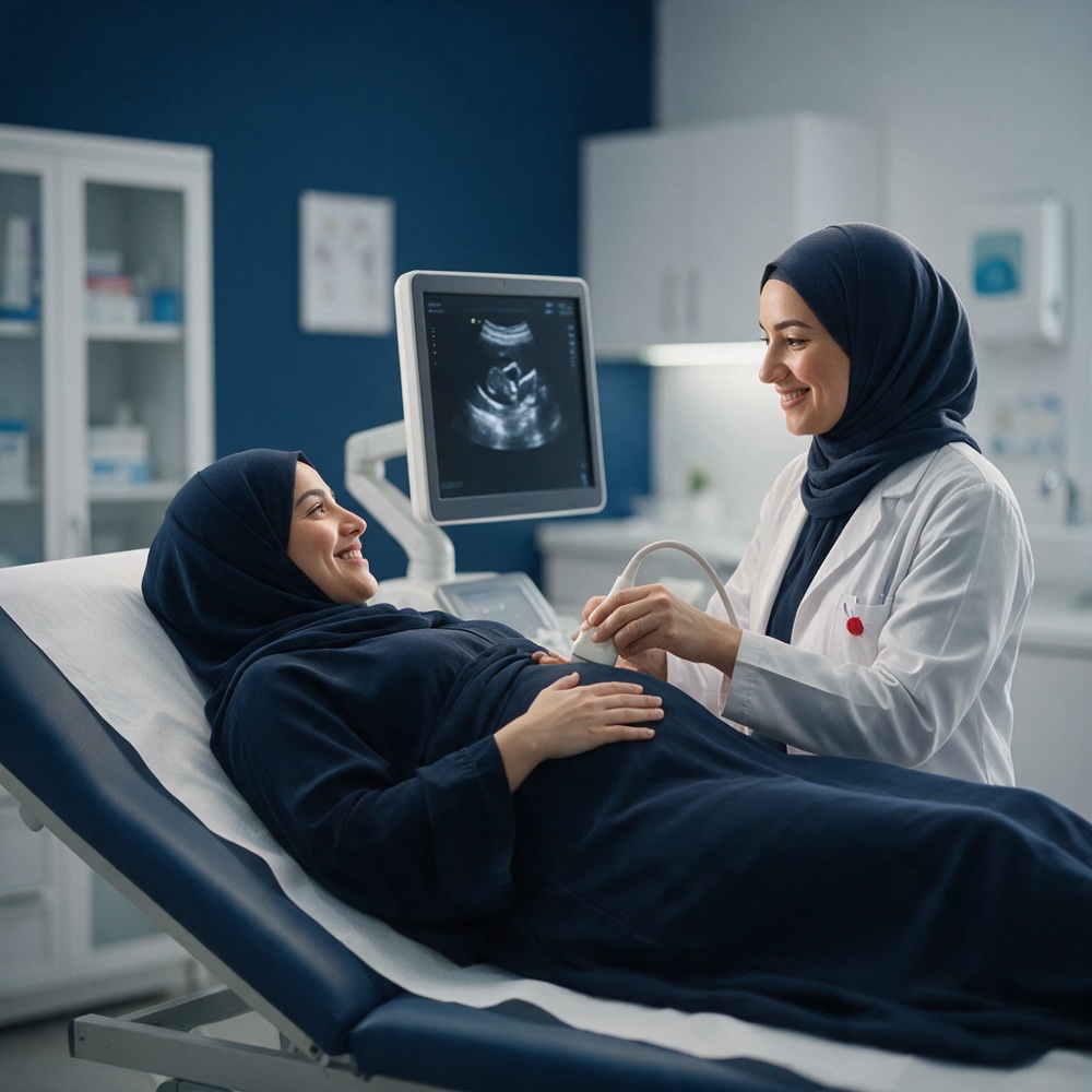
Obstetrics & gynecology clinic in Al-Taawun, Riyadh: Dr. Hanan Hilal — pregnancy follow-up and in-clinic ultrasound, detailed and 4D scans in the radiology department, NIPT, contraceptive implant and IUD, and pregnancy packages.
We are distinguished in the Medical World Clinics Polyclinic by having a group of distinguished doctors in all medical specialties